The Molecular Origins of Biological Color
How electronic structure shapes light absorption in pigments, proteins, and living tissues.
Sample article: use this as a formatting reference, then replace it with your own writing.
Color feels immediate. A leaf looks green; a carotenoid appears orange; blood looks red. Each pigment has a molecular structure that determines how it interacts with light.
Molecules do not absorb every wavelength equally
Electrons in atoms and molecules occupy allowed energy states. When incoming light carries an appropriate amount of energy, it can promote an electron from one state to another. Wavelengths that are absorbed are removed from the light that reaches us.
The color we perceive is therefore related to the light that remains.
Structure changes the possibilities
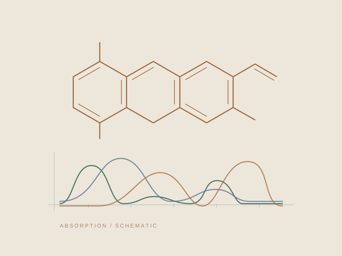
Change a molecule’s bonding pattern and you can change the spacing between its electronic energy levels. Conjugated systems are a familiar example: extending a network of alternating bonds can shift which wavelengths are absorbed.
In biological pigments, this connects electronic structure to function. Chlorophyll absorbs light that helps drive photosynthesis. The conjugated chain of a carotenoid absorbs visible light and contributes to its color.
A protein changes the environment
The behavior of a pigment also depends on its surroundings. A protein can position a chromophore, alter its chemical environment, and change how it interacts with light. The optical properties of a biological system belong to the molecule and its context.
What looks like a simple patch of color is the visible result of interactions happening at an electronic scale.